- HPI: 50M with acute chest pain
- Cath: PCI after cardiac arrest – LCX “subtotally stenosed” – peak hscTnT 2,696 ng/L
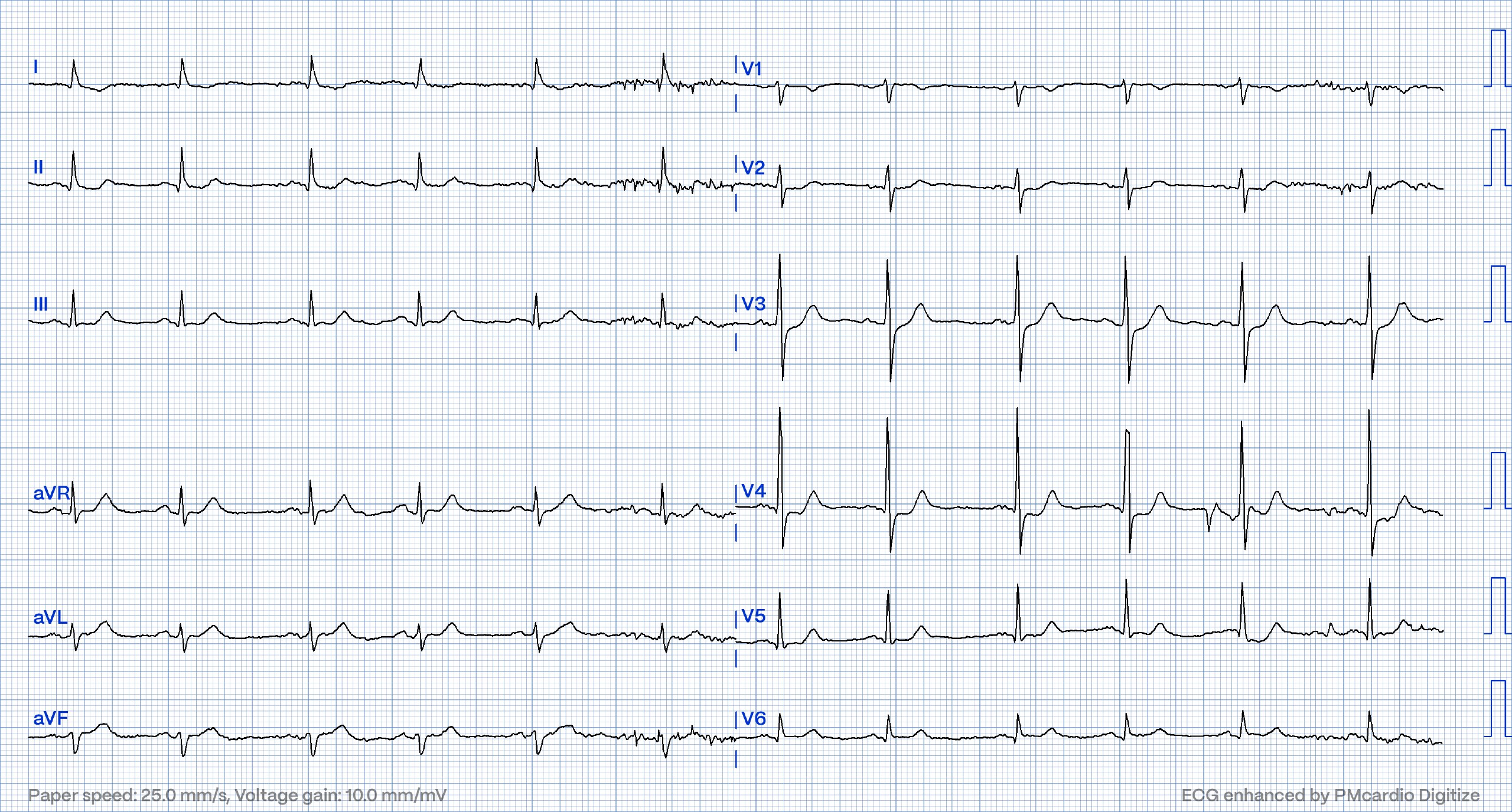
- ECG: Findings are specific for posterior (and also likely inferior) wall transmural acute infarction, most likely due to acute coronary occlusion (OMI). There is a relatively normal QRS yet there is STD maximal in V2-V4, which resolves from V4 to V6. The inferior leads may have a slightly full T wave (possibly hyperacute if compared to baseline which is unavailable), but there is no clear TWI in aVL
- Link to full case @ Dr. Smith’s ECG blog: 2023-06-21