- HPI: 45M with chest pain x 6 hr
- Cath: 100% proximal LAD occlusion
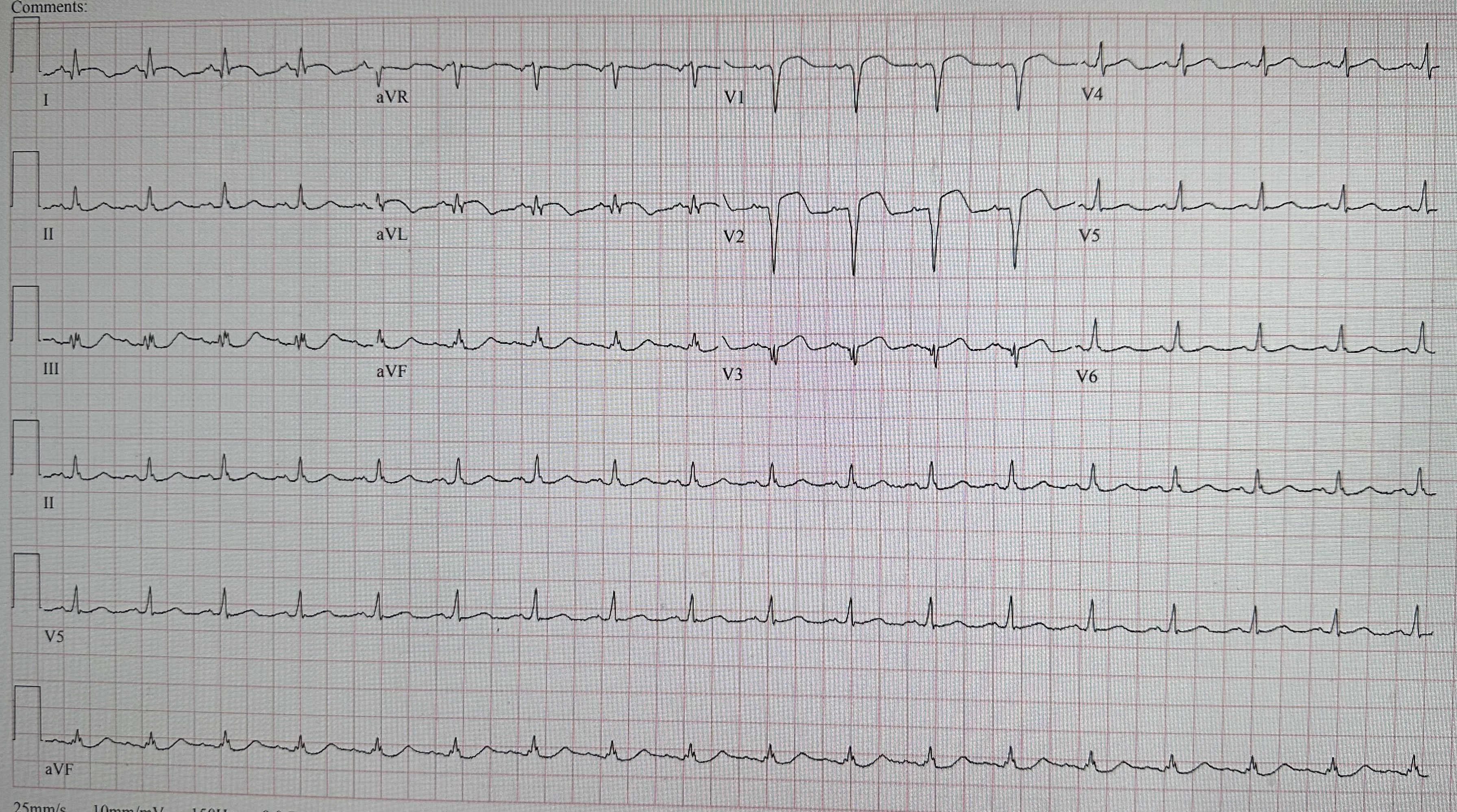
- ECG: 1)There is a QS-wave in V2. That indicates old or subacute MI. But the patient has no previous cardiac history, so we must assume it is new, and so subacute, and that is consistent with the 6-hour duration of pain. 2) The T-wave is also large for old MI. My rule is that if any lead has a T/QRS ratio >0.36, then it is acute OMI; if <0.36, then either old MI (LV aneurysm) or subacute OMI. 3) There is ST depression in V3 and V4. Always abnormal. 4) There is coving of the ST segment in I and aVL, with large inverted T-waves and reciprocally upright large inferior T-waves (these actually suggest some reperfusion, but as the patient has persistent symptoms, one must assume there is continued ischemia). 5) The proximal LAD will affect the territory of the LAD and of the 1st diagonal, which supplies the high lateral wall, resulting in the findings in I and aVL, and the reciprocal findings in III and aVF.
- Link to full case @ Dr. Smith’s ECG Blog: 2023-05-18