- HPI: 60F awoke with substernal chest pressure x 1/2 hour
- Cath: 99% thrombotic lesion in the proximal segment of a large obtuse marginal, with TIMI-2 flow
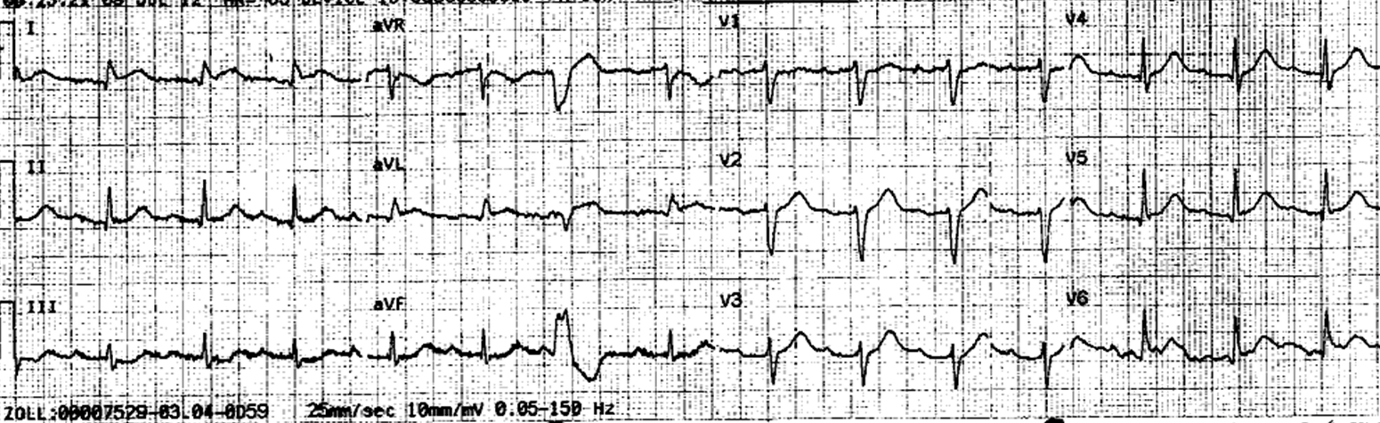
- Key ECG:There is sinus rhythm with one PVC. There is subtle ST elevation in aVL and I, with reciprocal ST depression in III and aVF, indicative of a circuflex (or obtuse marginal – OM – branch), or possibly and first diagonal, occlusion. There are also hyperacute T-waves in V4-V6, with some ST elevation, suggesting more widespread STEMI, such as LAD or very large OM or diagonal. The ST elevation is even easier to see in the PVC in lead aVL: it is discordant, as it should be, but out of proportion.
- Link to full case @ Dr. Smith’s ECG Blog: 2012-08-01