- HPI:60F w/ CAD s/p PCI presenting with chest pain x 3 hr.
- Cath: LAD thrombus
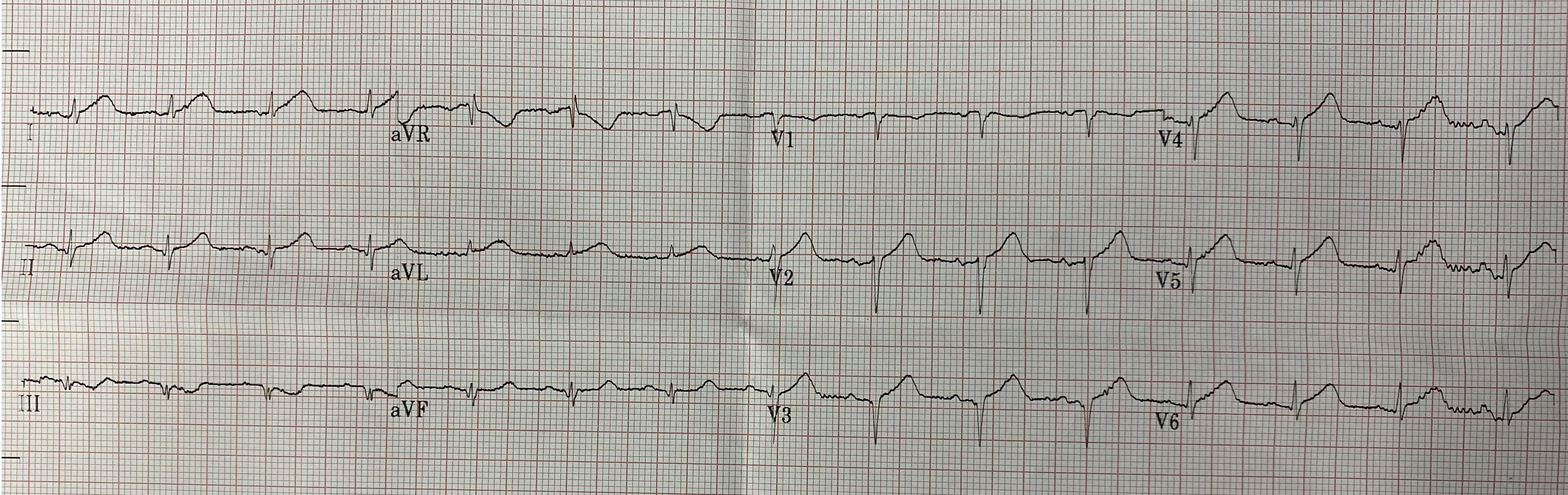
- Key ECG:There are hyperacute T-waves in I, aVL, V2-V6. These are wide, bulky, with large area under the curve relative to the QRS size. Furthermore, there is a QS-wave in V3 and qrS in V4, both suggestive of MI at some time (past or present). Dr. Smith has shown here and validated here that old MI has relatively small T-wave (by amplitude). In this situation (QS-waves), a T/QRS ratio >0.36 in any of V1-V4 is highly specific for ACUTE MI.
- Link to full case @ Dr. Smith’s ECG Blog: 2023-01-02