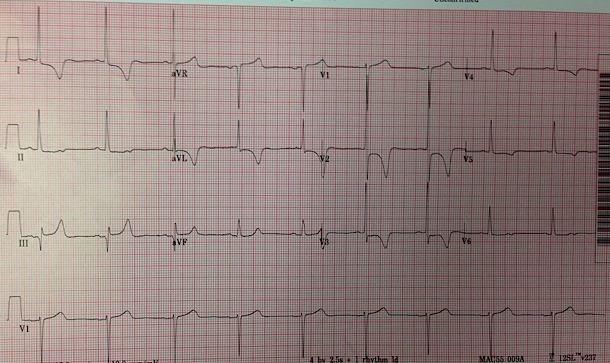
- HPI: Elderly woman with weakness. No chest pain
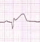
- Key ECG: One could mistake this for an inferior-posterior STEMI, as there is ST elevation in lead III with reciprocal ST depression in aVL, and there is ST depression in V2 and V3 with a tall R-wave (the mirror image of a posterior Q-wave), however the high voltage in I, V2, V3 and aVL meet “criteria” for LVH. ST elevation in III is a scooped-out saddleback, and that is because the ST segment is long and flat, and thus the T-wave is not hyperacute; rather, it has a narrow base. Contrast it with this wide-based T-wave in a true inferior STEMI. But doesn’t all ST elevation with reciprocal ST depression in aVL mean Acute MI? No! Rciprocal STD in aVL is frequently found in these STEMI mimics: LVH, LBBB, inferior LV aneurysm, myocarditis.
- Link to full case @ Dr. Smith’s ECG Blog: 2015-10-28