⚠️ Note:
- LV aneurysm should be suspected when there are QS-waves in any of leads V1-V4
- A QS-wave means a single negative deflection, without any R-wave or with only a tiny r-wave.
- Only about 70%-80% of patients with the ECG morphology of “LV aneurysm” actually have an LV aneurysm, as defined by echocardiographic dyskinesis.
- “LV aneurysm” is far less common in this era of reperfusion, in which STEMI is not allowed to progress to full infarction
- LV Aneurysm can be inferior, anterior, or posterior. Inferior aneurysm looks very much like acute MI because it does not get QS-waves, but rather QR-waves, which can also be present in acute MI.
🚨 Suggestive of OMI:
✅ Expected changes in LV Aneurysm:
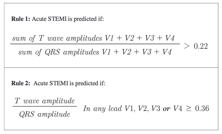
- In acute coronary occlusion, the T-wave is large, whereas in LV aneurysm, the T-wave is not so large. There are two pretty accurate formulas:
- T/QRS ratio < 0.36 in any of leads V1 – V4
- [TV1 + TV2 + TV3 + TV4] divided by [QRS V1 + QRS V2 + QRS V3 + QRS V4] < 0.22