- HPI: 60M with acute chest pain
- Cath: 100% thrombotic lesion of the proximal left circumflex (TIMI 0 flow)
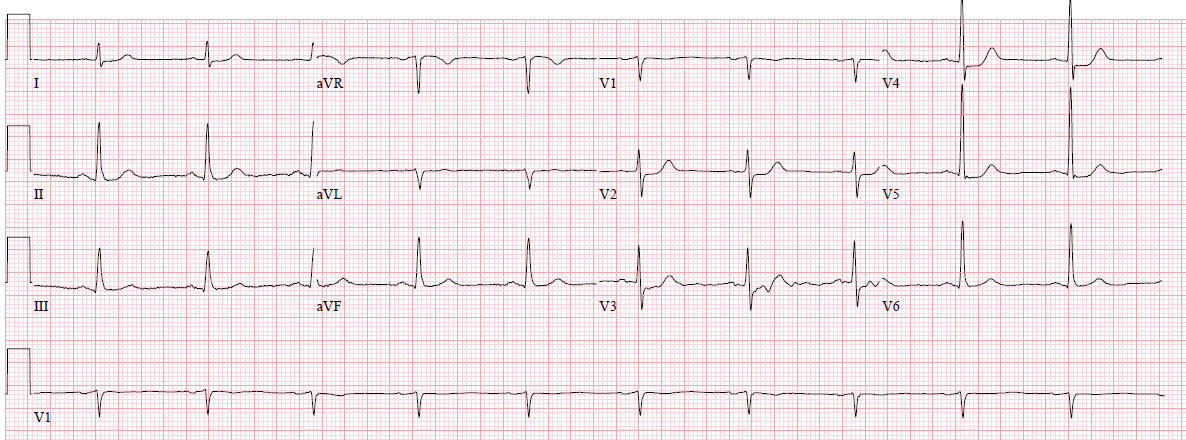
- Key ECG: There is sinus rhythm with normal QRS complex and ST depression in V2-V5, maximal in V3-V4. There is no ST depression in V6, II, III, or aVF, and no significant ST elevation in aVR, all confirming that the ST vector is not consistent with diffuse subendocardial ischemia, but rather a focal ST vector pointed at the posterior wall. It is posterior OMI until proven otherwise
- Link to full case @ Dr. Smith’s ECG Blog: 2019-02-16